Patient History
38-year-old man with gluteal abscess had undergone incision. Delayed healing and persistent secretion gave rise to the suspicion of an underlying fistula. Subtraction MR-Fistulography was performed to exclude the suspected diagnosis.
Image Findings
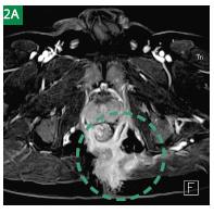
At coronal TIRM sequence high signal intensity abscess is shown within the left gluteus muscle (Fig. 1A) which communicates with a fluid-filled formation above the level of the levator muscle surrounding the lower rectum (Fig. 1B). Note also the reactive lymph nodes inside the mesorectum (Fig. 1A). On the axial contrast-enhanced 3D FLASH sequence after image subtraction a rectal fistula is clearly delimitable, forming a supralevatoric horseshoe (Fig. 2A). The internal opening is located at 9 o ’clock lithotomy position. The fistula passes the levator muscle at 4 o’clock (Fig. 2B) towards the fat of the left-sided ischiorectal fossa. The fistula retains pus and is directly connected to the residual gluteal abscess (Figs. 2C, D). MIP-reconstruction of the data derived from the 3D FLASH sequence is giving a survey of the whole extent of inflammation (Fig. 3).


Results and Discussions
To detect and precisely assess the extent of anorectal fistulas subtraction, MR-fistulography is extremely helpful. Contrastagent based, gradient-echo imaging provide excellent anatomic resolution and the capability to perform image reconstructions on the one hand as well as thin-sections and high sensitivity for inflammation on the other hand.

Fig. 3 3D FLASH, Maximum Intensity Projection (MIP) reconstruction.


