Patient History
48-year-old woman with cancer of the lower rectum received transanal endoscopic microsurgery (TEM) in 2003. In that year a recto-vaginal fistula was also closed. Currently, the patient presented with perianal pain and recurrent rectal cancer was diagnosed. Pelvic MRI was performed to determine the extent of the recurrent tumor.
Image Findings
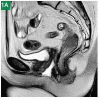
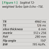
A sagittal T2-TSE sequence demonstrates the recurrent rectal cancer located above the anal canal infiltrating the rightsided levator muscle (Figs. 1A, B, C). Additionally, fluid is accumulated in the vaginal lumen after rectal water filling indicating ano-vaginal fistula. On the axial T2 SPACE sequence (Figs. 2A,B,C) and the corresponding axial contrast-enhanced VIBE sequence (Figs. 3A, B, C) the tumor is infiltrating the mesorectal fat, the mosrectal fascia and the parietal peritoneal fascia along a scar formation on the right side with small amounts of ascites indicative for peritoneal spread (Fig. 2A). Infiltration of the right levator muscle is seen (Figs. 2,3). There are no lymph nodes present.


Results and Discussions
Pelvic anatomy can be imaged exquisitely with the 3D, highresolution T2 SPACE sequence as well as with the high-resolution contrast-enhanced T1 VIBE sequence. A combination of both appears to be useful particularly for local staging of rectal cancer.