Chronic pulmonary diseases (CPD) represent a major global health burden and are the leading cause of death among lung diseases [1]. Each year, more than 55 million new cases are diagnosed worldwide. Approximately 10% of patients affected by CPDs are children*.
CPDs comprise a spectrum of diseases that require continuous surveillance. In this context, assessment of lung function, particularly perfusion (blood flow within the lungs) and ventilation (air flow through the lungs), is of central importance. Nuclear medicine is currently considered the the gold standard for functional lung imaging.
However, when repeated follow-up imaging is required, radiation exposure has to be taken into account, especially in vulnerable groups such as pediatric* patients.
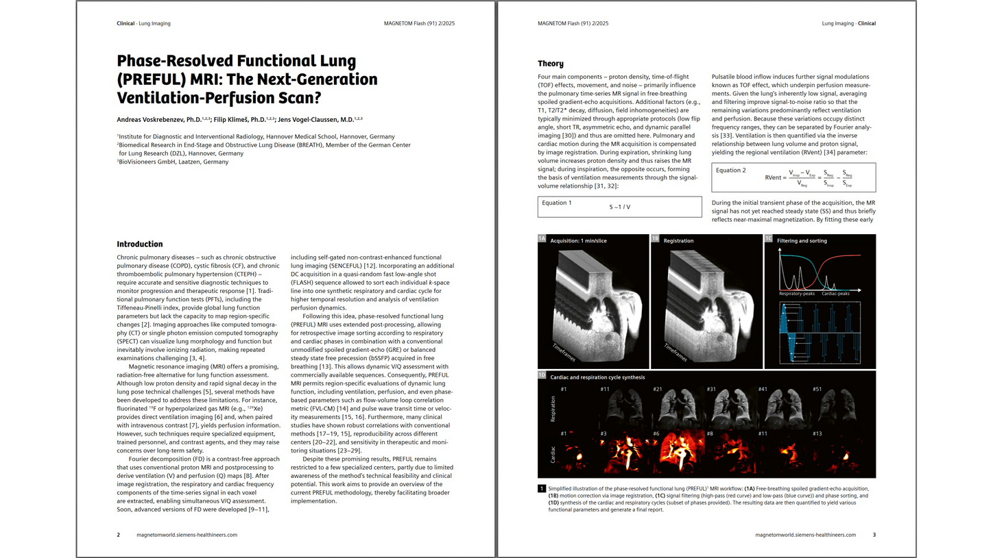
LungMaps is a new proton MRI-based application that provides functional lung information without the need for tracers, contrast agents, or ionizing radiation. It is based on the PREFUL method [2], which has been clinically evaluated across a wide range of patient populations. Although LungMaps is a post-processing application, it relies on a 2D MRI acquisition protocol with sufficient temporal resolution to capture both respiratory- and cardiac-based signal variations in the lungs.
Protocols for 0.55T, 1.5T, and 3T MRI systems are available below and serve as input data for LungMaps.
- Download 0.55T MAGNETOM Free (syngo MR XA80) LungMaps protocol (.exar1 & PDF) (zip) 0.47MB (binary/octet-stream) 0.17 MB
- Download 1.5T MAGNETOM Sola (syngo MR XA61) LungMaps protocol (.exar1 & PDF) (zip) 0.53MB (binary/octet-stream) 0.15 MB
- Download 3T MAGNETOM Vida (syngo MR XA60) LungMaps protocol (.exar1 & PDF) (zip) 0.41MB (binary/octet-stream) 0.15 MB
References
[1] Global Burden of Disease Collaborative Network. Global Burden of Disease Study 2021 (GBD 2021) Burden and Strength of Evidence by Risk Factor 1990-2021. Seattle, USA: Institute for Health Metrics and Evaluation (IHME), 2024. https://www.thelancet.com/gbd
[2] Voskrebenzev A, et al.: Feasibility of Quantitative Regional Ventilation and Perfusion Mapping With Phase-Resolved Functional Lung (PREFUL) MRI in Healthy Volunteers and COPD, CTEPH, and CF Patients; Magn Reson Med 79:2306-2314 (2018) https://doi.org/10.1002/mrm.26893
*MR scanning has not been established as safe for imaging fetuses and infants less than two years of age. The responsible physician must evaluate the benefits of the MR examination compared to those of other imaging procedures.
Learn more:
Phase-Resolved Functional Lung (PREFUL) MRI: The Next-Generation Ventilation-Perfusion Scan?
Andreas Voskrebenzev, Ph.D.1,2,3; Filip Klimeš, Ph.D.1,2,3; Jens Vogel-Claussen, M.D.1,2,3
1 Institute for Diagnostic and Interventional Radiology, Hannover Medical School, Hannover, Germany
2 Biomedical Research in End-Stage and Obstructive Lung Disease (BREATH), Member of the German Center
for Lung Research (DZL), Hannover, Germany
3 BioVisioneers GmbH, Laatzen, Germany
Contact
Andreas Voskrebenzev, Ph.D.
Department of Radiology,
Charité Universitätsmedizin Berlin,
corporate member of Freie Universität Berlin
and Humboldt-Universität zu Berlin
Charitéplatz 1
10117 Berlin
Germany
https://radiologie.charite.de/metas/kontakt_aufnehmen_mit/adresse/dr_rer_nat_andreas_voskrebenzev