Neurodegenerative Diseases
Neurodegenerative Diseases (NDD) are a huge global burden. These conditions affect more than 60 million people worldwide and are responsible for more than 2 million fatalities every year[1]. Among these diseases, Alzheimer’s Disease (AD), Parkinson’s Disease (PD), and Multiple Sclerosis (MS) are most widely known and the most relevant ones in the context of magnetic resonance imaging.
Alzheimer’s Disease (AD)
AD has seen a high interest since 2021 when the first anti-amyloid drug was released by the FDA. As of today, these so-called disease modifying therapies (DMT[2]) are seen as a beacon of hope for AD patients who could only receive medication addressing the symptoms so far. Side effects of DMTs are so called Amyloid-Related Imaging Abnormalities (ARIA) which require follow-up imaging with MRI in defined intervals[3-5]. The protocols can be found here:
Protocols

ASNR-Recommended AD Therapeutic Imaging Protocols
Tammie L.S. Benzinger, M.D., Ph.D.; et al.
Washington University School of Medicine in St. Louis, Mallinckrodt Institute of Radiology, St. Louis, MO, USA
Articles
A clinician’s guide to detecting and diagnosing ARIA in Alzheimer’s disease patients

Within-Subject Comparison of Brain Acquisition Protocols with 16- and 32-Channel Coils on a Hybrid PET-MR Scanner
Maria Eugenia Caligiuri, Ph.D.; et al. (University Magna Graecia, Catanzaro, Italy)
Assessing Brain Volumes Using MorphoBox Prototype
Alexis Roche1,2,3; Bénédicte Maréchal1,2,3; Tobias Kober 1,2,3; Gunnar Krueger 4 ; Patric Hagmann1 ;
Philippe Maeder 1 ; Reto Meuli 1
1 Department of Radiology, University Hospital (CHUV), Lausanne, Switzerland
2 Advanced Clinical Imaging Technology, Siemens Healthineers, Lausanne, Switzerland
3 Signal Processing Laboratory (LTS 5), École Polytechnique Fédérale de Lausanne, Lausanne, Switzerland
4 Siemens Medical Solutions USA, Boston, MA
Presentations
Dementia Imaging Update: Role of the Radiologist in the Era of Disease-Modifying Therapies
Ana M. Franceschi, MD, PhD
(Lenox Hill Hospital, Donald and Barbara Zucker School of Medicine at Hofstra/Northwell, Hempstead, NY, USA)
ECR Lunch Symposium 2025
Vienna, Austra
ARIA Imaging and Workflows
Petrice Cogswell, M.D., Ph.D. (Mayo Clinic, Rochester, MN, USA)
Siemens Healthineers Symposium RSNA 2024
Parkinson’s Disease (PD)
PD is well known for its symptoms covering resting tremor, rigor (muscle stiffness), bradykinesia (slowness of movement) and ataxia (balance problems)[6]. Clinical research with MRI on PD has seen progress, in particular in the field of Quantitative Susceptibility Mapping (QSM), an emerging advanced MR method relying on T2*-weighted gradient echo imaging. Here you can find more information on PD and QSM:
Articles
Insights from 7T: GRE T2*-weighted Sequences Uncover Biomarkers in Neurological Diseases
Jing Jing1,2; Zhe Zhang1; Decai Tian2; Tao Feng2; Hua Pan2; Yuan Li3; Xiaoye Wang3; Jianxun Qu3
1Tiantan Neuroimaging Center of Excellence, China National Clinical Research Center for Neurological Diseases, Beijing Tiantan Hospital, Capital Medical University, Beijing, China
2Department of Neurology, Beijing Tiantan Hospital, Capital Medical University, Beijing, China
3Siemens Healthineers, Beijing, China
Utility of Quantitative Susceptibility Mapping in Neuroradiology: Initial Clinical Experience at a Tertiary Referral Hospital
Yangsean Choi, M.D., Ph.D.1; Sugil Kim, Ph.D.2
1Department of Radiology, Asan Medical Center, Seoul, Republic of Korea
2MR Research Collaboration, Siemens Healthineers Ltd, Seoul, Republic of Korea
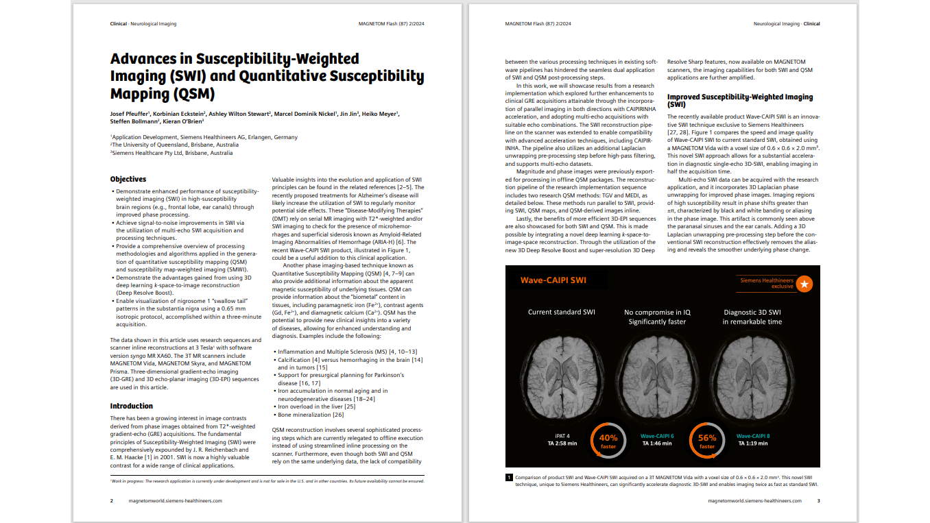
Advances in Susceptibility-Weighted Imaging (SWI) and Quantitative Susceptibility Mapping (QSM)
Josef Pfeuffer1, Korbinian Eckstein2, Ashley Wilton Stewart2, Marcel Dominik Nickel1, Jin Jin3, Heiko Meyer1, Steffen Bollmann2, Kieran O‘Brien3
1Application Development, Siemens Healthineers AG, Erlangen, Germany
2The University of Queensland, Brisbane, Australia
3Siemens Healthcare Pty Ltd, Brisbane, Australia
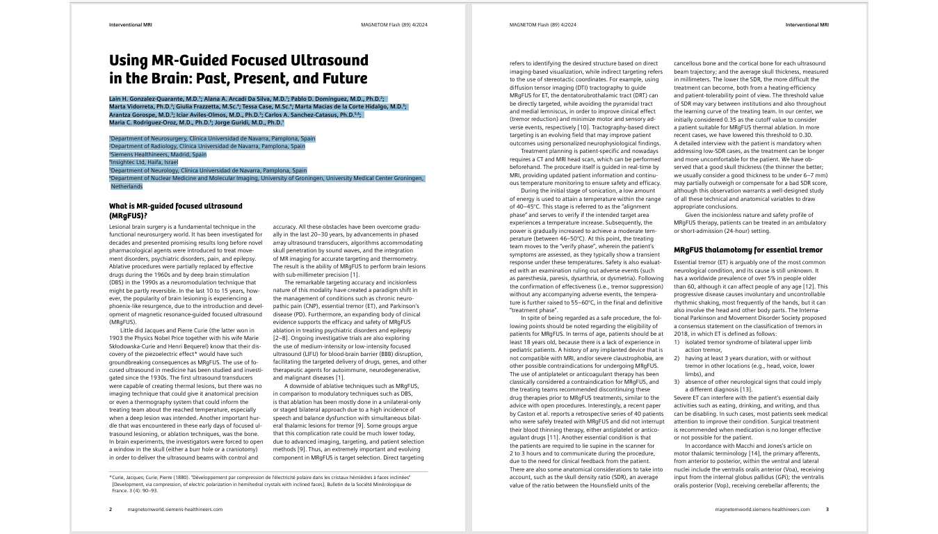
Using MR-Guided Focused Ultrasound in the Brain: Past, Present, and Future
Lain H. Gonzalez-Quarante, M.D.1; Alana A. Arcadi Da Silva, M.D.1; Pablo D. Domínguez, M.D., Ph.D.2; Marta Vidorreta, Ph.D.3; Giulia Frazzetta, M.Sc.4; Tessa Case, M.Sc.4; Marta Macias de la Corte Hidalgo, M.D.5; Arantza Gorospe, M.D.5; Iciar Aviles-Olmos, M.D., Ph.D.5; Carlos A. Sanchez-Catasus, Ph.D.5,6; Maria C. Rodriguez-Oroz, M.D., Ph.D.5; Jorge Guridi, M.D., Ph.D.1<
1Department of Neurosurgery, Clínica Universidad de Navarra, Pamplona, Spain
2Department of Radiology, Clínica Universidad de Navarra, Pamplona, Spain
3Siemens Healthineers, Madrid, Spain
4Insightec Ltd, Haifa, Israel
5Department of Neurology, Clínica Universidad de Navarra, Pamplona, Spain
6 Department of Nuclear Medicine and Molecular Imaging, University of Groningen, University Medical Center Groningen, Netherlands
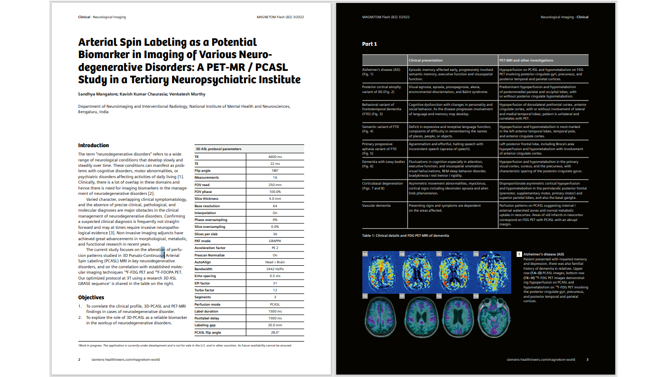
Arterial Spin Labeling as a Potential Biomarker in Imaging of Various Neuro degenerative Disorders: A PET-MR / PCASL Study in a Tertiary Neuropsychiatric Institute
Sandhya Mangalore; Kavish Kumar Chaurasia; Venkatesh Murthy
Department of Neuroimaging and Interventional Radiology, National Institute of Mental Health and Neurosciences, Bengaluru, India
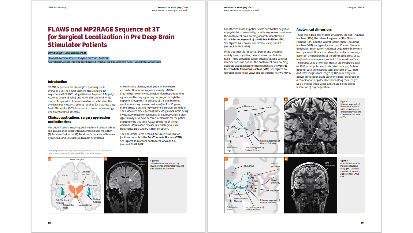
FLAWS and MP2RAGE Sequence at 3T for Surgical Localization in Pre Deep Brain Stimulator Patients
David Shipp1 ; Tobias Kober, Ph.D.2
1Monash Medical Centre, Clayton, Victoria, Australia
2 Advanced Clinical Imaging Technology, Siemens Medical Solutions-CIBM, Lausanne, Switzerland
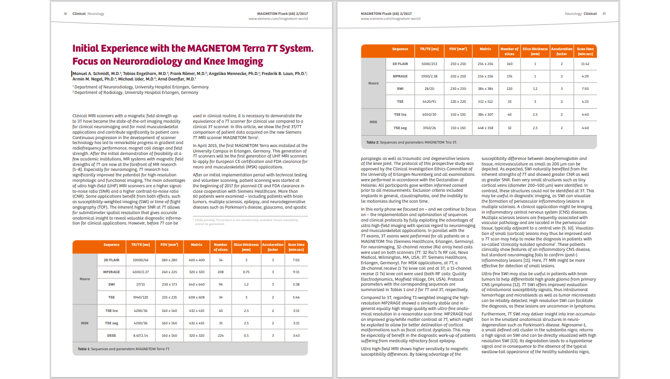
Initial Experience with the MAGNETOM Terra 7T System. Focus on Neuroradiology and Knee Imaging
Manuel A. Schmidt, M.D.1 ; Tobias Engelhorn, M.D.1 ; Frank Römer, M.D.2 ; Angelika Mennecke, Ph.D.1 ; Frederik B. Laun, Ph.D.2 ; Armin M. Nagel, Ph.D.2 ; Michael Uder, M.D.2 ; Arnd Doerfler, M.D.1
1Department of Neuroradiology, University Hospital Erlangen, Germany
2Department of Radiology, University Hospital Erlangen, Germany
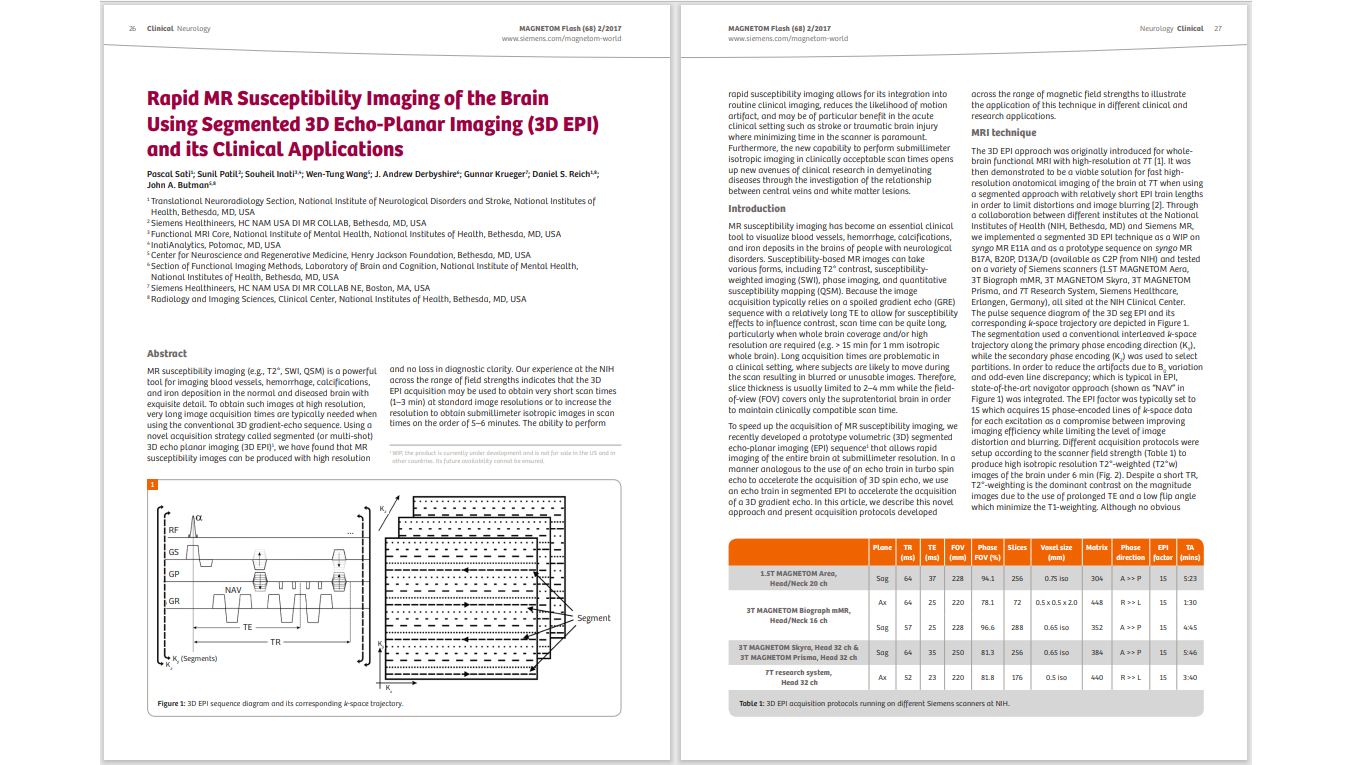
Rapid MR Susceptibility Imaging of the Brain Using Segmented 3D Echo-Planar Imaging (3D EPI) and its Clinical Applications
Pascal Sati1 ; Sunil Patil2 ; Souheil Inati3,4; Wen-Tung Wang5 ; J. Andrew Derbyshire6 ; Gunnar Krueger7 ; Daniel S. Reich1,8; John A. Butman5,8
1 Translational Neuroradiology Section, National Institute of Neurological Disorders and Stroke, National Institutes of Health, Bethesda, MD, USA
2 Siemens Healthineers, HC NAM USA DI MR COLLAB, Bethesda, MD, US
3 Functional MRI Core, National Institute of Mental Health, National Institutes of Health, Bethesda, MD, USA
4 InatiAnalytics, Potomac, MD, USA
5 Center for Neuroscience and Regenerative Medicine, Henry Jackson Foundation, Bethesda, MD, USA
6 Section of Functional Imaging Methods, Laboratory of Brain and Cognition, National Institute of Mental Health, National Institutes of Health, Bethesda, MD, USA
7 Siemens Healthineers, HC NAM USA DI MR COLLAB NE, Boston, MA, USA
8Radiology and Imaging Sciences, Clinical Center, National Institutes of Health, Bethesda, MD, USA
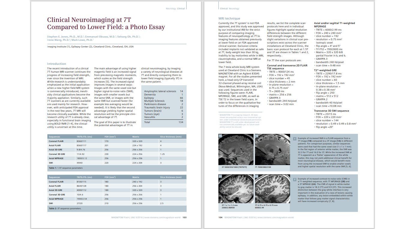
Clinical Neuroimaging at 7T Compared to Lower Field: a Photo Essay
Stephen E. Jones, Ph.D., M.D.1 ; Emmanuel Obusez, M.D.1 ; Sehong Oh, Ph.D.1 ; Irene Wang, Ph.D.2 ; Mark Lowe, Ph.D.1
Cleveland Clinic, Cleveland, OH, USA
1 Imaging Institute
2 Epilepsy Center
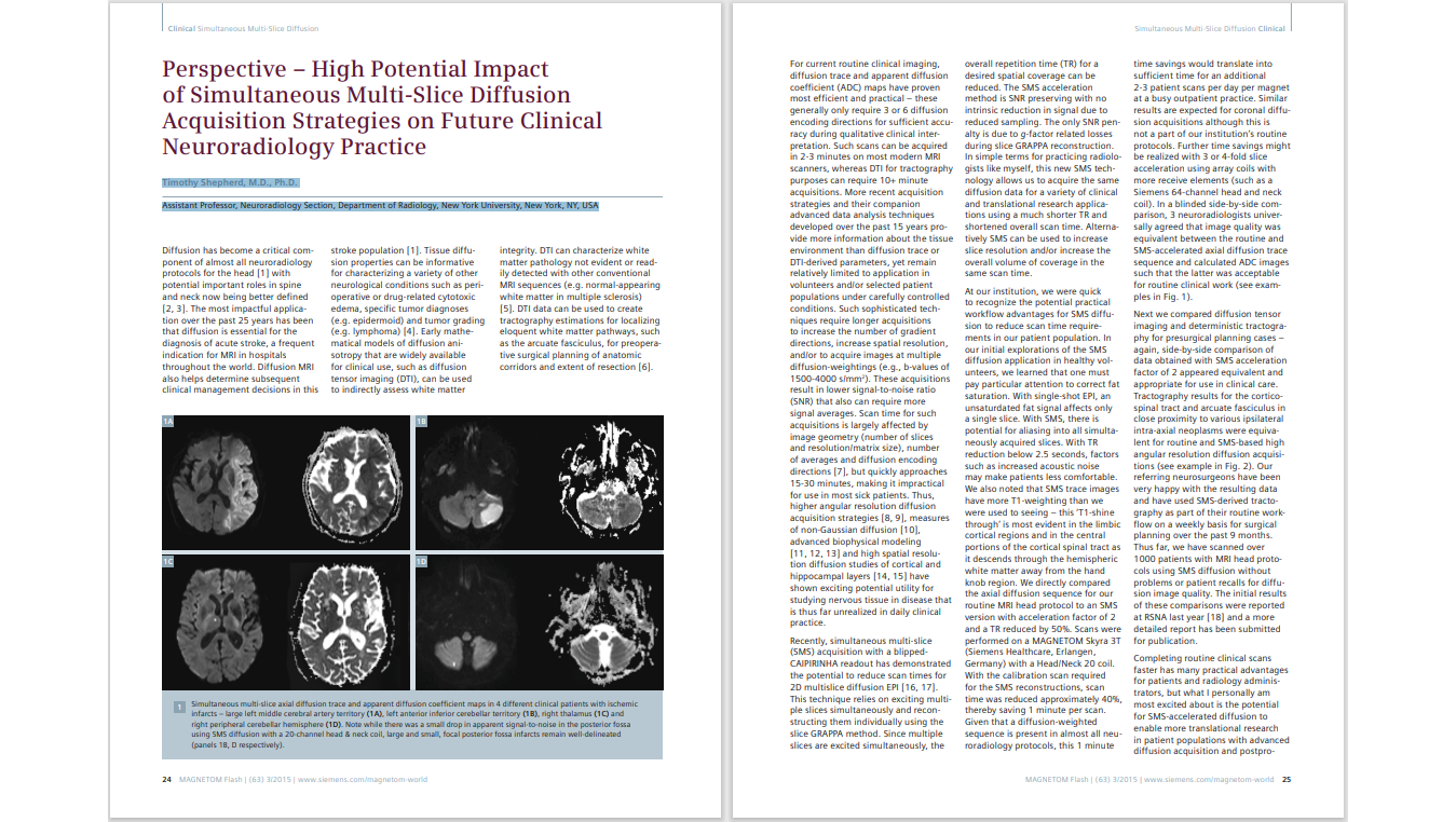
Perspective – High Potential Impact of Simultaneous Multi-Slice Diffusion Acquisition Strategies on Future Clinical Neuroradiology Practice
Timothy Shepherd, M.D., Ph.D. Assistant Professor, Neuroradiology Section, Department of Radiology, New York University, New York, NY, USA
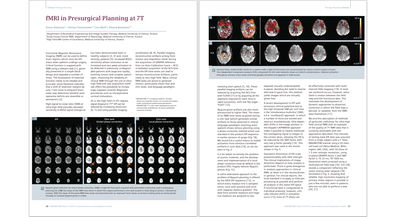
fMRI in Presurgical Planning at 7T
Simon Robinson1,3; Florian Fischmeister2,3; Eva Matt2,3; Roland Beisteiner2,3
1 Department of Biomedical Engineering and Image-Guided Therapy, Medical University of Vienna, Austria
2 Study Group Clinical fMRI, Department of Neurology, Medical University of Vienna, Austria
3High Field MR Center of Excellence, Medical University of Vienna, Austria
Multiple Sclerosis (MS)
MS is one of the few NDDs where MRI is the primary imaging modality for diagnosis. The diagnostic guidelines are the McDonald criteria, and they have seen their latest update in 2024. Here you can find MS related publications as well as protocols that follow the McDonald criteria[7].
Protocols
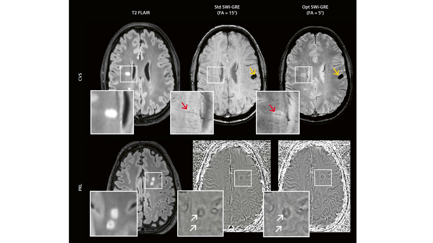
Optimized Susceptibility MRI Sequences for Novel Diagnostic Biomarkers of Multiple Sclerosis
Sreekanth Madhusoodhanan Nair, Ph.D.; Nader Binesh, Ph.D.; Marcel Maya, M.D.; Pascal Sati, Ph.D.
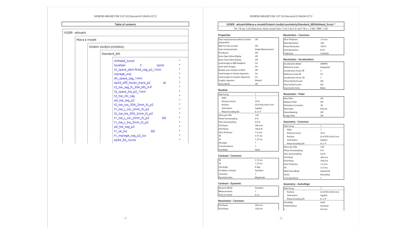
Diagnostic, Monitoring, and Safety MRI Protocols for Multiple Sclerosis
Based on the 2021 and 2024 MAGNIMS Consensus Recommendations and the McDonald Criteria
Manuela Vaněčková, Ph.D., MBA
General University Hospital in Prague, Department of Magnetic Resonance (MRI) of the Radiodiagnostic Clinic at the Neurological Clinic, Prague, Czech Republic
Articles
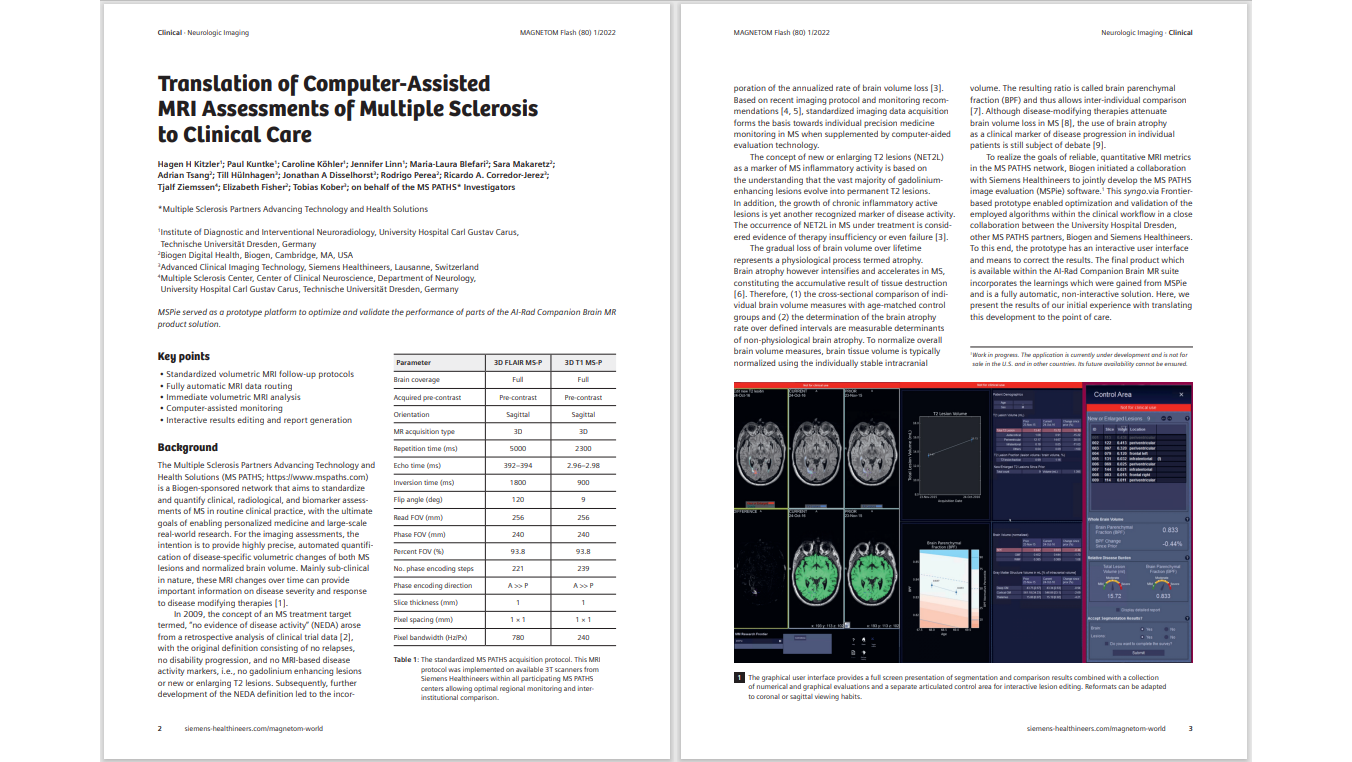
Translation of Computer-Assisted MRI Assessments of Multiple Sclerosis to Clinical Care
Hagen H Kitzler1; Paul Kuntke1; Caroline Köhler1; Jennifer Linn1; Maria-Laura Blefari2; Sara Makaretz2; Adrian Tsang2; Till Hülnhagen3; Jonathan A Disselhorst3; Rodrigo Perea2; Ricardo A. Corredor-Jerez3; Tjalf Ziemssen4; Elizabeth Fisher2; Tobias Kober3
1Institute of Diagnostic and Interventional Neuroradiology, University Hospital Carl Gustav Carus, Technische Universität Dresden, Germany
2Biogen Digital Health, Biogen, Cambridge, MA, USA
3Advanced Clinical Imaging Technology, Siemens Healthineers, Lausanne, Switzerland
4Multiple Sclerosis Center, Center of Clinical Neuroscience, Department of Neurology, University Hospital Carl Gustav Carus, Technische Universität Dresden, Germany
Advanced Diffusion MRI in Neurological Diseases: The Multiple Sclerosis Model
Simona Schiavi, Ph.D.1; Matilde Inglese, M.D., Ph.D.1,2
1Department of Neuroscience, Rehabilitation, Ophthalmology, Genetics, Maternal and Child Health (DINOGMI), University of Genoa, Italy
2IRCCS Ospedale Policlinico San Martino, Genoa, Italy
Update on FLAIR Fusion in Multiple Sclerosis Follow-up and Beyond: An Indispensable Tool in Clinical Routine
Andreas Bartsch, M.D.1; Thomas Illigen2
1Radiologie Bamberg, Germany
2Siemens Healthineers, Erlangen, Germany
This is an update on the article “FLAIR Fusion in Multiple Sclerosis Follow-Up: An Indispensable Tool in Clinical Routine” by Stéphane Cantin and Emeline Lamain (Groupe Clinique du Mail, Grenoble, France), and Thomas Troalen and Melisa Bakir (Siemens Healthineers, Saint-Denis, France) that was published in MAGNETOM Flash (69) 3/2017.
The Next Generation – Advanced Design Low-field MR Systems
Val M. Runge, M.D.; Johannes T. Heverhagen, M.D., Ph.D.
Department of Diagnostic, Interventional and Pediatric Radiology, University Hospital of Bern, Inselspital, University of Bern, Switzerland
Iterative Denoising Applied to 3D SPACE CAIPIRINHA: A New Approach to Accelerate 3D Brain Examination in Clinical Routine
Alexis Vaussy1; Michael Eliezer2; Nicolas Menjot de Champfleur3; Emmanuelle Le Bars3; Thien Huong Nguyen4; Christophe Habas4; Alto Stemmer5; Thomas Troalen1
1Siemens Healthineers, Saint-Denis, France
2Department of Neuroradiology, Lariboisière University Hospital, Paris, France
3Department of Neuroradiology, Hôpital Gui de Chauliac, Montpellier University Medical Center, Montpellier, France
4Radiodiagnostic and Medical Imaging, Quinze-Vingts National Ophthalmology Hospital, Paris, France 5Siemens Healthineers, Erlangen, Germany
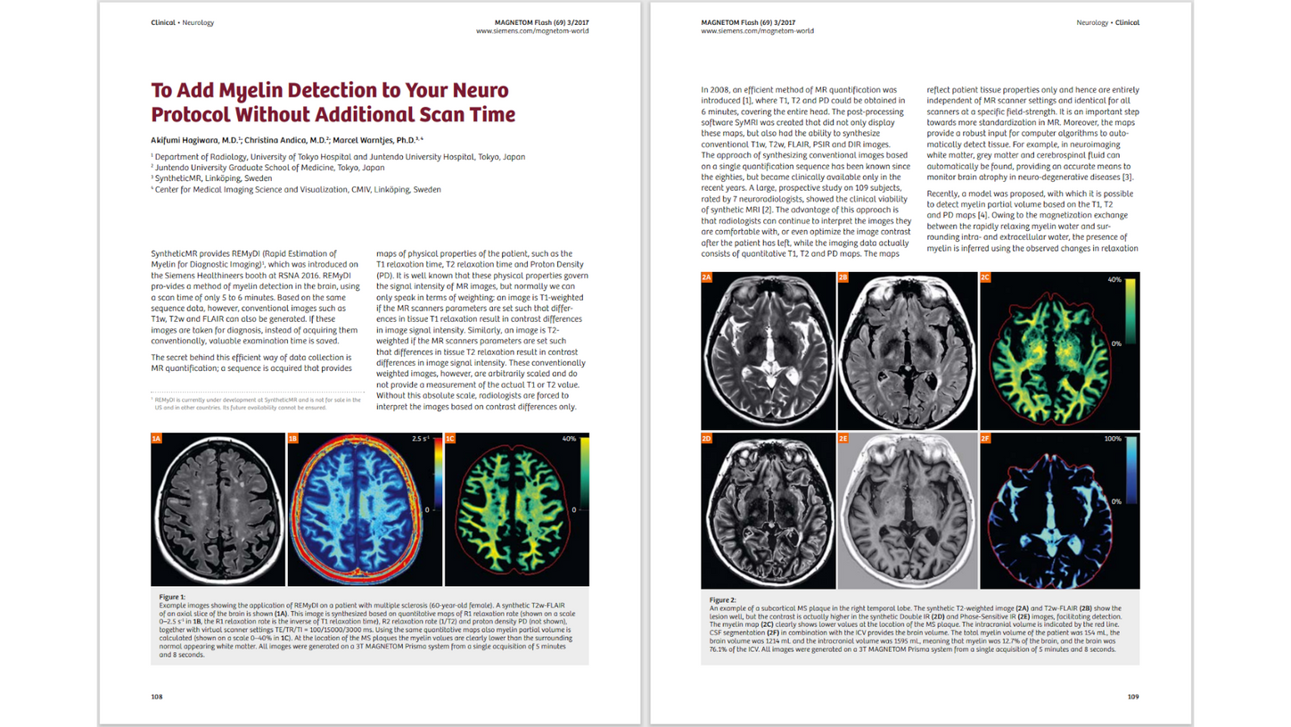
To Add Myelin Detection to Your Neuro Protocol Without Additional Scan Time
Akifumi Hagiwara, M.D.1; Christina Andica, M.D.2; Marcel Warntjes, Ph.D.3, 4
1Department of Radiology, University of Tokyo Hospital and Juntendo University Hospital, Tokyo, Japan
2Juntendo University Graduate School of Medicine, Tokyo, Japan
3SyntheticMR, Linköping, Sweden
4Center for Medical Imaging Science and Visualization, CMIV, Linköping, Sweden
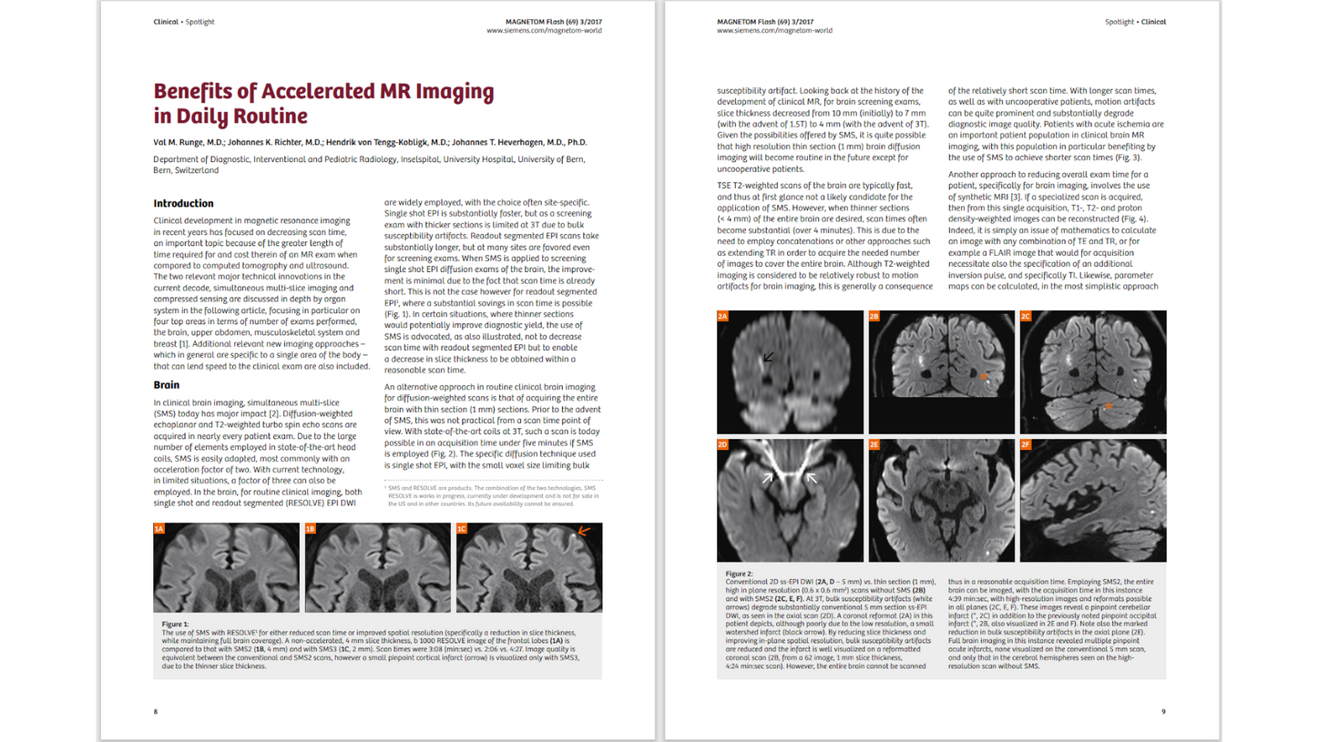
Benefits of Accelerated MR Imaging in Daily Routine
Val M. Runge, M.D.; Johannes K. Richter, M.D.; Hendrik von Tengg-Kobligk, M.D.; Johannes T. Heverhagen, M.D., Ph.D.
Department of Diagnostic, Interventional and Pediatric Radiology, Inselspital, University Hospital, University of Bern, Bern, Switzerland
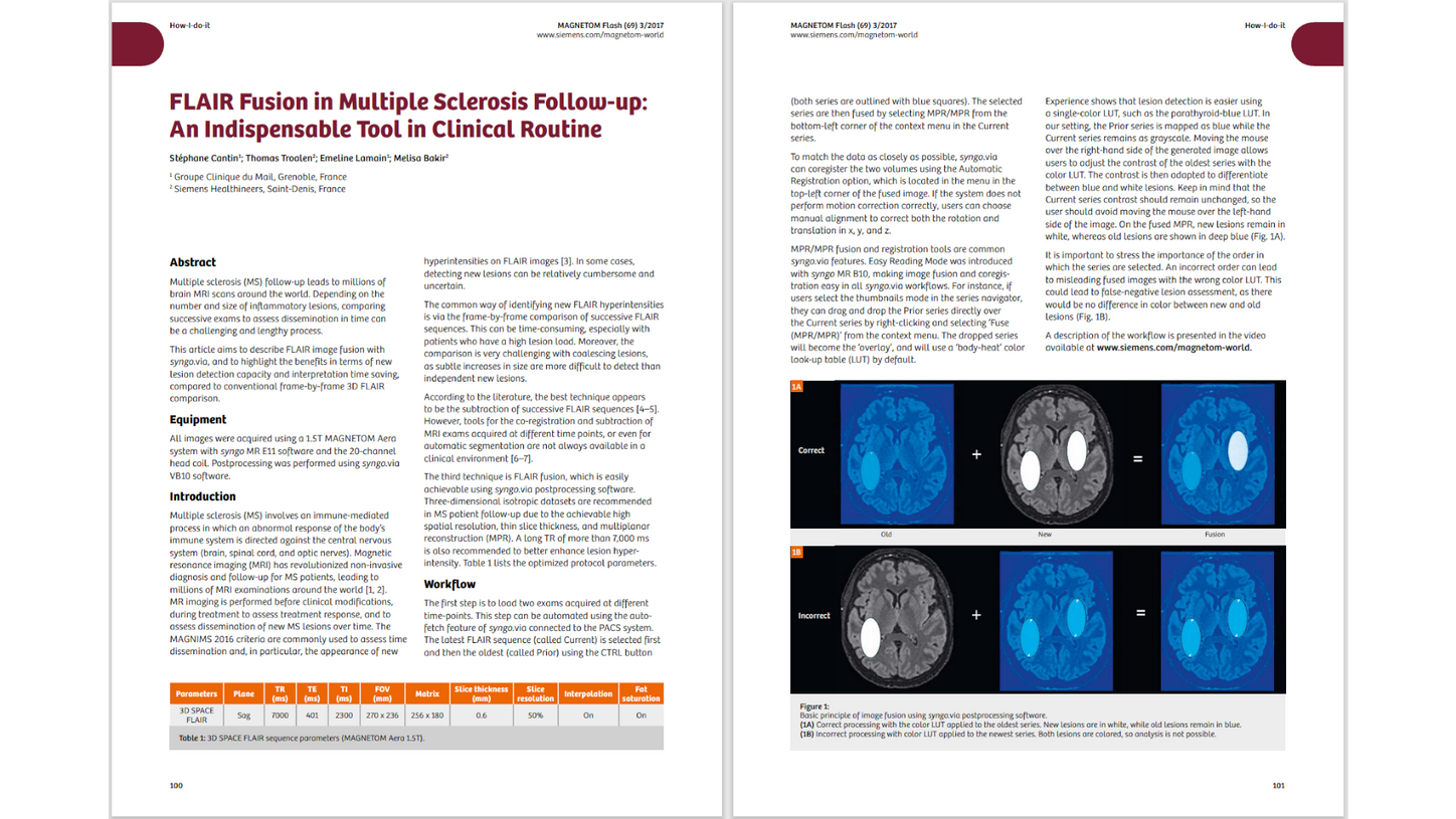
FLAIR Fusion in Multiple Sclerosis Follow-up: An Indispensable Tool in Clinical Routine
Stéphane Cantin1; Thomas Troalen2; Emeline Lamain1; Melisa Bakir2
1Groupe Clinique du Mail, Grenoble, France
2Siemens Healthineers, Saint-Denis, France
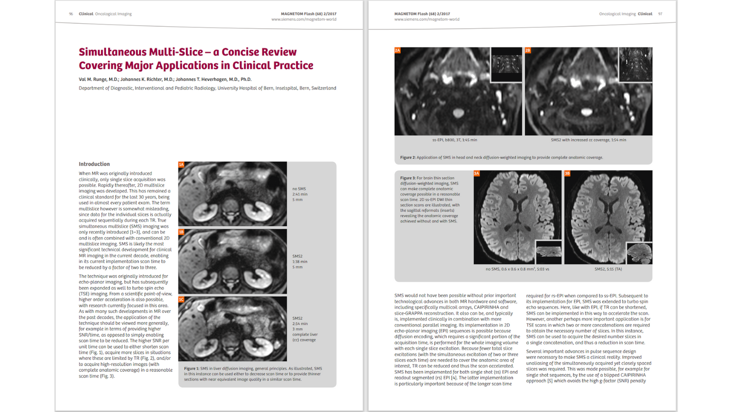
Simultaneous Multi-Slice – a Concise Review Covering Major Applications in Clinical Practice
Val M. Runge, M.D.; Johannes K. Richter, M.D.; Johannes T. Heverhagen, M.D., Ph.D.
Department of Diagnostic, Interventional and Pediatric Radiology, University Hospital of Bern, Inselspital, Bern, Switzerland
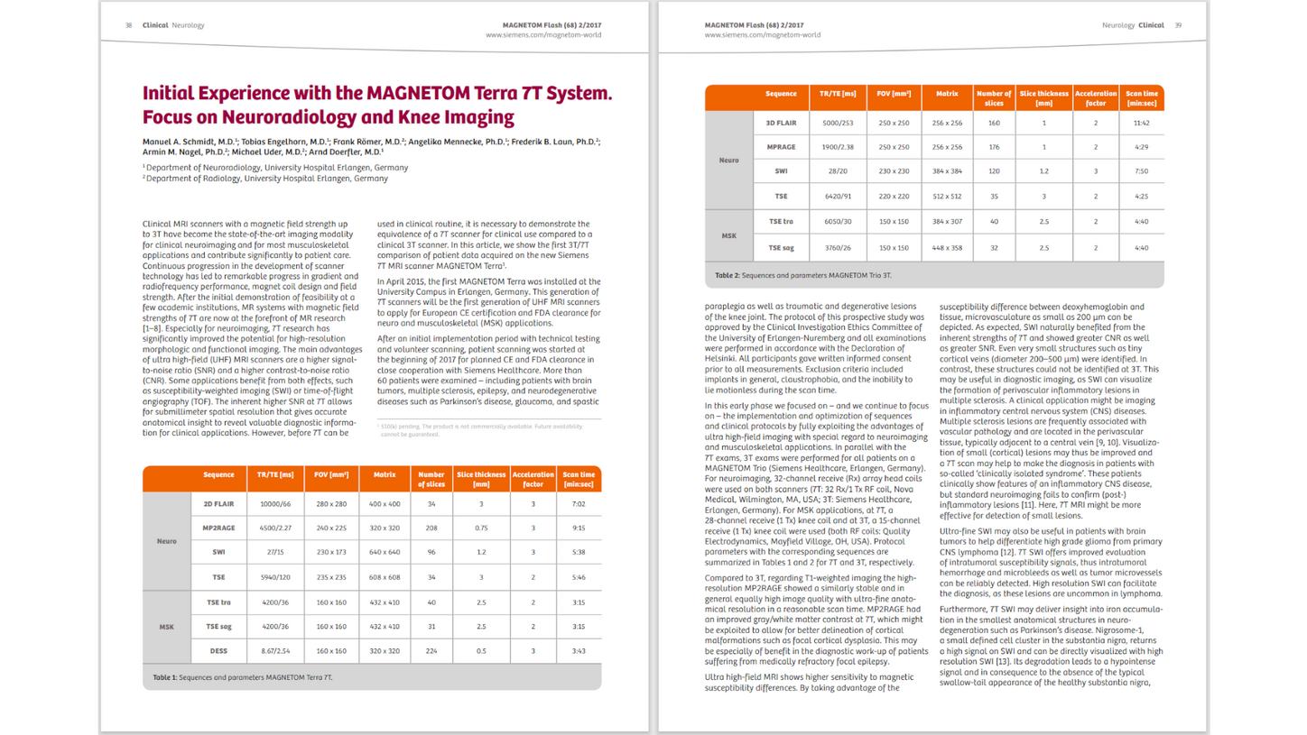
Initial Experience with the MAGNETOM Terra 7T System. Focus on Neuroradiology and Knee Imaging
Manuel A. Schmidt, M.D.1; Tobias Engelhorn, M.D.1; Frank Römer, M.D.2; Angelika Mennecke, Ph.D.1; Frederik B. Laun, Ph.D.2; Armin M. Nagel, Ph.D.2; Michael Uder, M.D.2; Arnd Doerfler, M.D.1
1Department of Neuroradiology, University Hospital Erlangen, Germany
2Department of Radiology, University Hospital Erlangen, Germany
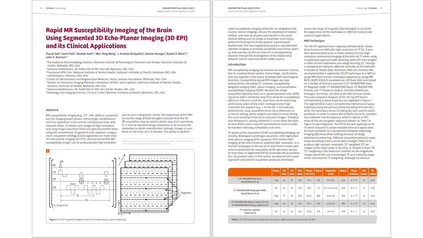
Rapid MR Susceptibility Imaging of the Brain Using Segmented 3D Echo-Planar Imaging (3D EPI) and its Clinical Applications
Pascal Sati1; Sunil Patil2; Souheil Inati3,4; Wen-Tung Wang5; J. Andrew Derbyshire6; Gunnar Krueger7; Daniel S. Reich1,8; John A. Butman5,8
1Translational Neuroradiology Section, National Institute of Neurological Disorders and Stroke, National Institutes of Health, Bethesda, MD, USA
2Siemens Healthineers, HC NAM USA DI MR COLLAB, Bethesda, MD, USA
3Functional MRI Core, National Institute of Mental Health, National Institutes of Health, Bethesda, MD, USA
4InatiAnalytics, Potomac, MD, USA
5Center for Neuroscience and Regenerative Medicine, Henry Jackson Foundation, Bethesda, MD, USA
6Section of Functional Imaging Methods, Laboratory of Brain and Cognition, National Institute of Mental Health, National Institutes of Health, Bethesda, MD, USA
7Siemens Healthineers, HC NAM USA DI MR COLLAB NE, Boston, MA, USA 8Radiology and Imaging Sciences, Clinical Center, National Institutes of Health, Bethesda, MD, USA
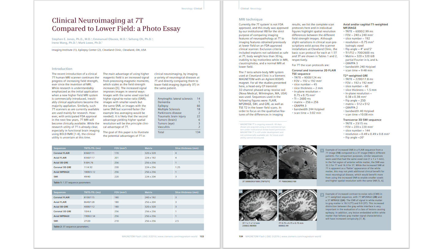
Clinical Neuroimaging at 7T Compared to Lower Field: a Photo Essay
Stephen E. Jones, Ph.D., M.D.1; Emmanuel Obusez, M.D.1; Sehong Oh, Ph.D.1; Irene Wang, Ph.D.2; Mark Lowe, Ph.D.1
Cleveland Clinic, Cleveland, OH, USA
1Imaging Institute
2Epilepsy Center
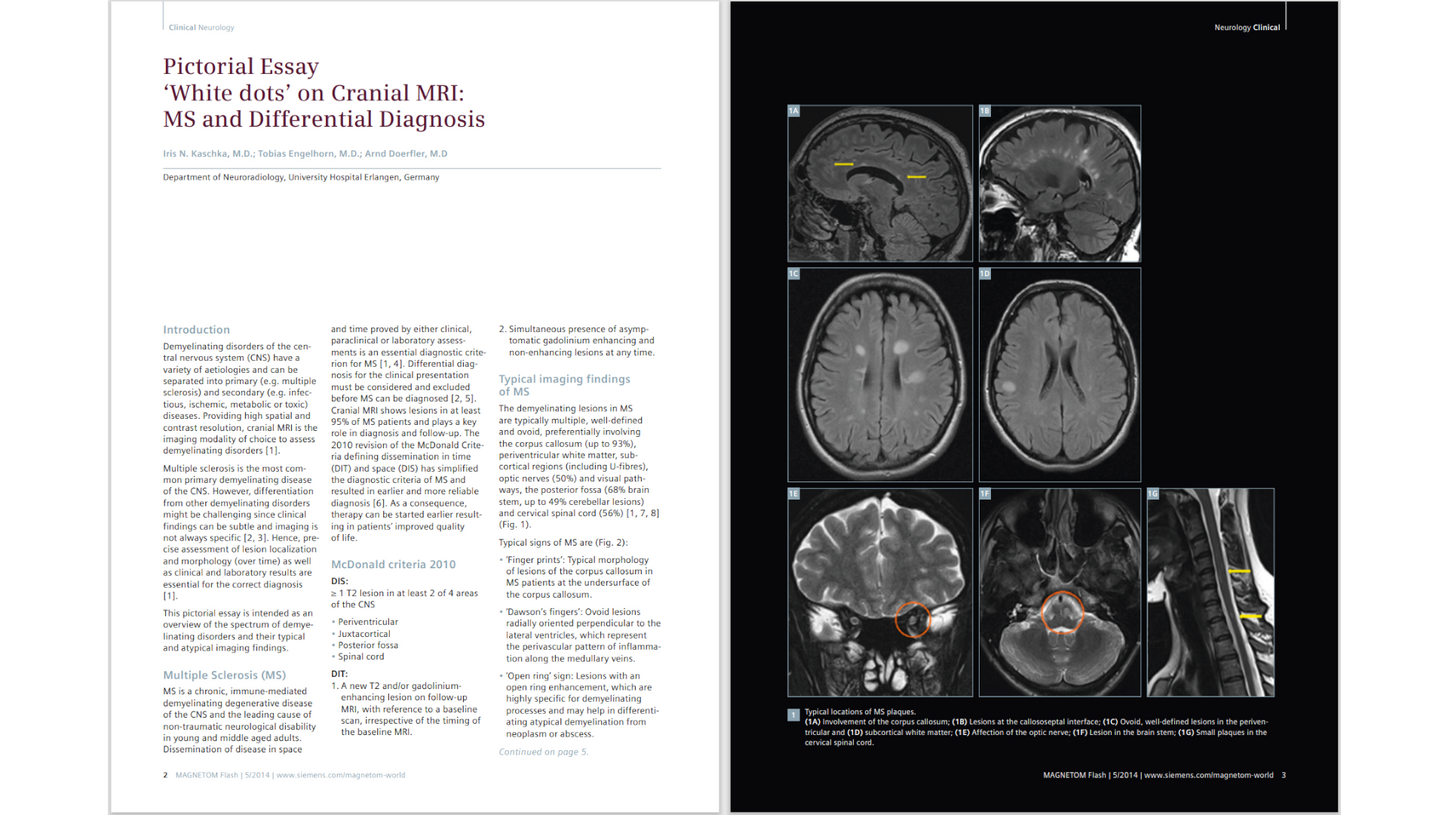
Pictorial Essay ‘White dots’ on Cranial MRI: MS and Differential Diagnosis
Iris N. Kaschka, M.D.; Tobias Engelhorn, M.D.; Arnd Doerfler, M.D.
Department of Neuroradiology, University Hospital Erlangen, Germany
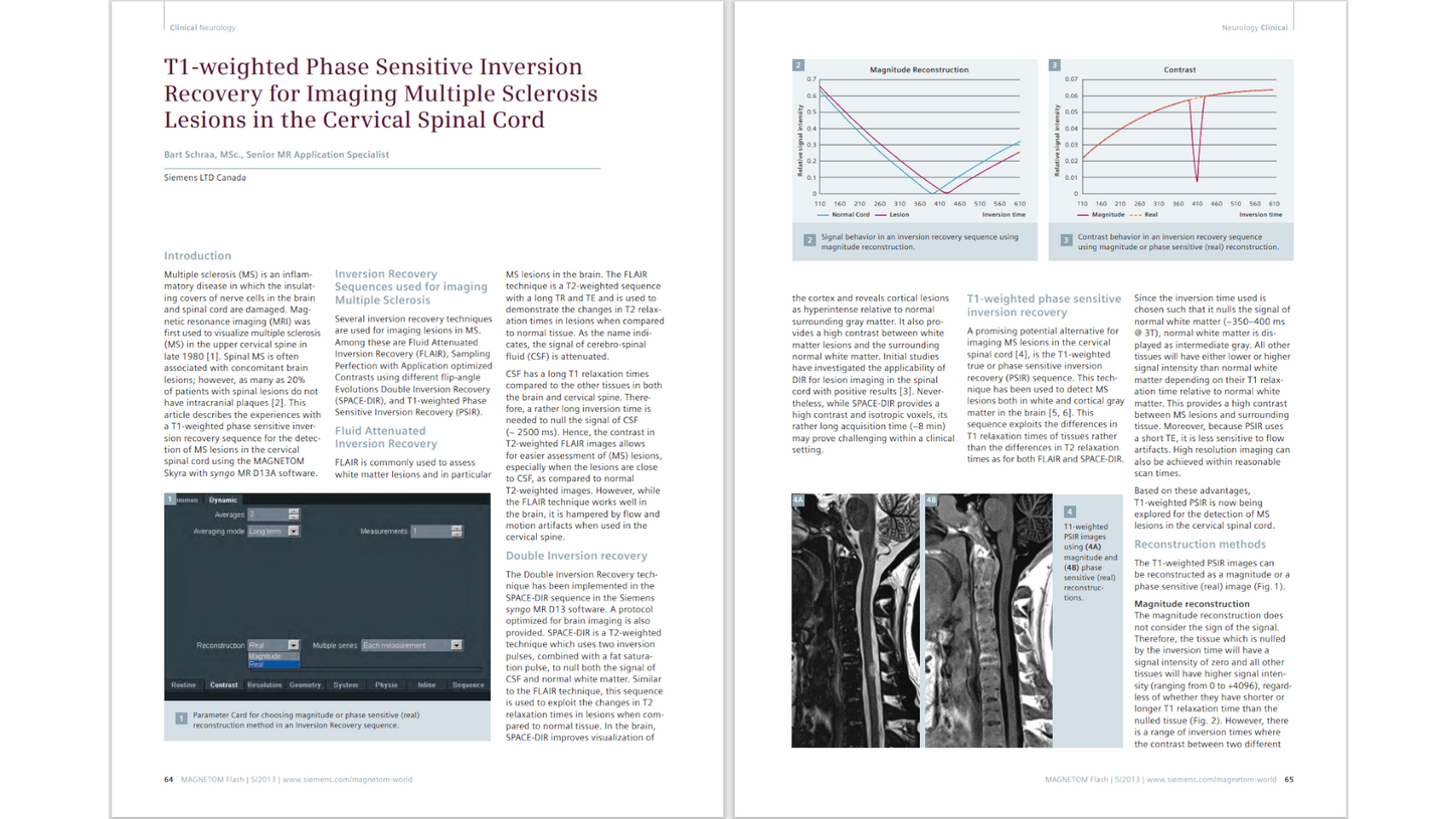
T1-weighted Phase Sensitive Inversion Recovery for Imaging Multiple Sclerosis Lesions in the Cervical Spinal Cord
Bart Schraa, MSc., Senior MR Application Specialist
Siemens LTD Canada
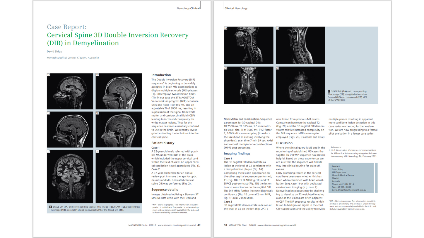
Case Report: Cervical Spine 3D Double Inversion Recovery (DIR) in Demyelination
David Shipp
Monash Medical Centre, Clayton, Australia
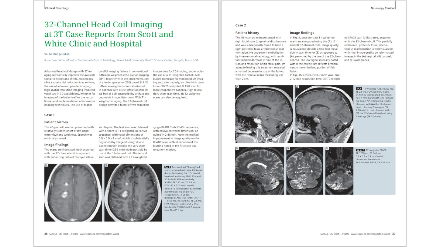
32-Channel Head Coil Imaging at 3T Case Reports from Scott and White Clinic and Hospital
Val M. Runge, M.D.
Robert and Alma Moreton Centennial Chair in Radiology, Texas A&M University Health Science Center, Temple, Texas, USA
Presentations
MRI Techniques in Multiple Sclerosis: New Diagnostic and Research Developments
Matilde Inglese, M.D., Ph.D.
University of Genoa, Italy / Icahn School of Medicine at Mount Sinai, New York, NY, USA
11th MAGNETOM World Summit
Wave CAIPI for Routine Clinical Brain MRI
Susie Huang, M.D., Ph.D.
Athinoula A. Martinos Center for Biomedical Imaging, Massachusetts General Hospital, Boston, MA, USA
11th MAGNETOM World Summit
Clinical translation: From 0 to 1500 patients in one year
Srinivasan Mukundan, M.D., Ph.D. (Brigham & Women’s Hospital, Boston, MA, USA)
Brain and spine: What to expect next from 7T?
Maxime Guye, M.D., Ph.D. (CRMBM, Aix-Marseille University, Marseille, France)